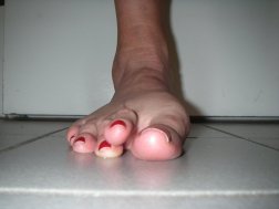
What Is Mortons Neuroma

Overview

interdigital neuromaMorton?s Neuroma is a pathological condition of the common digital nerve in the foot, most frequently between the third and fourth metatarsals (third inter-metatarsal space). The nerve sheath becomes abnormally thickened with fibrous (scar) tissue and the nerve fibres eventually deteriorate.This condition is named for the American surgeon, Thomas George Morton (1835-1903), who first recognised the condition in 1876. Incidentally his father was the dentist who discovered the anaesthetics; initially Nitrous oxide, the very gas used today in cryosurgery for the condition his son lent his name to? Morton?s neuroma.

Causes

The exact cause is as yet unclear. However there are a number of theories. Some expert s believe problems with the design of the foot makes some people more prone to Morton?s neuroma. Having flat feet or a high arch for example encourages the foot to slide forwards which can put excess pressure on the metatarsals. Bunions and hammer toes also increase the likelihood of developing Morton?s. However simply wearing high heels or any form of tight shoes that put pressure on the bones in the feet can also lead to a Morton?s . Typically the condition comes on between the age of 40 and 50. It is far more common in women than men – three out of four sufferers are women.

Symptoms

Patients with a Morton’s neuroma typically experience a sharp, shooting or burning pain, usually at the base of the forefoot or toes, which radiates into the two affected toes. Sometimes the pain may also radiate into the foot. The pain is often associated with the presence of pins and needles and numbness.

Diagnosis

Based on the physical examination, your doctor usually can diagnose a Morton’s neuroma without additional testing. A foot X-ray may be ordered to make sure that there isn’t a stress fracture, but it will not show the actual neuroma. If the diagnosis is in doubt, your doctor may request magnetic resonance imaging (MRI) of the foot.

Non Surgical Treatment

The most important factor in the treatment of Morton’s neuroma is changing footwear. Sometimes a cushioned dome pad can be worn inside the shoe and this helps spread the metatarsal heads and decrease pressure on the nerve. There are other products that can be worn between the toes with certain types of shoes or when the client is barefoot. These toe spacers will help reverse biomechanical patterns that aggravate the nerve compression. Massage can be helpful, but should not be performed with deep pressure between the metatarsal heads. Additional pressure in this region can aggravate the nerve compression and prolong the pathology.

Surgical Treatment

If other therapies have not worked it may be necessary to perform surgery. As surgery may result in permanent numbness in the affected toe, doctors ten to use this procedure as a last resort. However, in most cases surgery is extremely effective. The patient usually receives a local anesthetic. Surgery involves either removing the nerve, or removing the pressure on the nerve. Two surgical approaches are possible. The dorsal approach, the surgeon makes an incision on the top of the foot, allowing the patient to walk soon after surgery, because the stitches are not on the weight-bearing side of the foot. The plantar approach, the surgeon makes an incision on the sole of the foot. In most cases the patient will be in crutches for about three weeks. The resulting scar may make walking uncomfortable. However, with this approach the neuroma can be reached easily and resected without cutting any structures. There is a small risk of infection around the toes after surgery.

How You Can Prevent Inferior Calcaneal Spur

Inferior Calcaneal Spur

Overview

Heel spurs are new bone that forms in response to stress to the heel. They serve to protect the bone against the development of microfractures. Spurs start out as cartilage and progress to solid bone. They are present in about 50 percent of the population, yet not everyone has heel pain- that’s the first clue that heel spurs don’t always cause heel pain.

Causes

Early signs of heel pain are usually due to plantar fasciitis, the inflammation of the plantar fascia. It is probably the most common cause of heel pain seen by the podiatrist. It is seen in all groups of people; runners, athletes, weekend warriors, people who have jobs requiring a fair amount of standing, walking, or lifting, and those who have recently gained weight. The pain most often manifests itself after periods of non-weight bearing when the plantar fascia is given a chance to rest, so your first steps cause a sudden strain to the tissue. Pain is most common with the first steps in the morning and after periods of rest. Fortunately, upwards of 80 percent of those treated early do remarkably well with conservative therapy.

Calcaneal Spur

Symptoms

Heel spurs may or may not cause symptoms. Symptoms are usually related to the plantar fasciitis. You may experience significant pain. Your heel pain may be worse in the morning when you first wake up or during certain activities.

Diagnosis

Your doctor, when diagnosing and treating this condition will need an x-ray and sometimes a gait analysis to ascertain the exact cause of this condition. If you have pain in the bottom of your foot and you do not have diabetes or a vascular problem, some of the over-the-counter anti-inflammatory products such as Advil or Ibuprofin are helpful in eradicating the pain. Pain creams, such as Neuro-eze, BioFreeze & Boswella Cream can help to relieve pain and help increase circulation.

Non Surgical Treatment

Heel spurs can be solved with simple solutions that do not involve surgery. Avoiding extended periods of activity such as running, sports and walking. Applying ice directly to the heel for 5 minutes at a time. This helps soothe and reduce inflammation. Lose weight to reduce stress on your heels. A series of simple exercises. Inexpensive orthotic shoe inserts. The best way to treat heel spurs is by treating the underlying cause of the problem. This involves correcting the dynamics of your foot motion with orthotic insoles.

Surgical Treatment

When chronic heel pain fails to respond to conservative treatment, surgical treatment may be necessary. Heel surgery can provide relief of pain and restore mobility. The type of procedure used is based on examination and usually consists of releasing the excessive tightness of the plantar fascia, called a plantar fascia release. Depending on the presence of excess bony build up, the procedure may or may not include removal of heel spurs. Similar to other surgical interventions, there are various modifications and surgical enhancements regarding surgery of the heel.

What Are The Signs And Symptoms Of Posterior Calcaneal Spur

Posterior Calcaneal Spur

Overview

Heel spurs are a common foot problem resulting from excess bone growth on the heel bone. The bone growth is usually located on the underside of the heel bone, extending forward to the toes. One explanation for this excess production of bone is a painful tearing of the plantar fascia connected between the toes and heel. This can result in either a heel spur or an inflammation of the plantar fascia, medically termed plantar fascitis. Because this condition is often correlated to a decrease in the arch of the foot, it is more prevalent after the age of six to eight years, when the arch is fully developed.

Causes

Some causes of heel spurs include abnormal or lopsided walking, which places excessive stress on the heel bone, ligaments and nerves Running, jogging or jumping, especially on hard surfaces. Poorly fitted or badly worn shoes, especially those lacking appropriate arch support, excess weight and obesity.

Calcaneal Spur

Symptoms

Heel spurs result in a jabbing or aching sensation on or under the heel bone. The pain is often worst when you first arise in the morning and get to your feet. You may also experience pain when standing up after prolonged periods of sitting, such as work sessions at a desk or car rides. The discomfort may lessen after you spend several minutes walking, only to return later. Heel spurs can cause intermittent or chronic pain.

Diagnosis

Sharp pain localized to the heel may be all a doctor needs to understand in order to diagnose the presence of heel spurs. However, you may also be sent to a radiologist for X-rays to confirm the presence of heel spurs.

Non Surgical Treatment

There are many ways to treat heel spurs. Some remedies you can even do at home once a podiatrist shows you how. Heel spur treatment is very similar to treatment of plantar fasciitis. Here are a few of the most common treatments. First, your doctor will assess which activities are causing your symptoms and suggest rest and time off from these activities. Ice packs are used to control pain and reduce symptoms. Certain exercises and stretches help you to feel relief quickly. Medications that reduce inflammation and decrease pain are also used. Sometimes cortisone injections are given. Often special shoe orthotics can help to take the pressure off of the plantar fascia and reduce symptoms. Night splints that keep your heel stretched are sometimes recommended. Rarely, surgery is an option. A new treatment called extracorporeal shock wave therapy is being studied.

Surgical Treatment

When chronic heel pain fails to respond to conservative treatment, surgical treatment may be necessary. Heel surgery can provide pain relief and restore mobility. The type of procedure used is based on examination and usually consists of releasing the excessive tightness of the plantar fascia, called a plantar fascia release. The procedure may also include removal of heel spurs.

Prevention

Walk around before you buy shoes. Before you purchase your shoes, do the following. Re-lace the shoes if you’re trying on athletic shoes. Start at the farthest eyelets and apply even pressure to the laces as you come closer to the tongue of the shoe. Make sure that you can wiggle your toes freely inside of the shoe. Also, make sure that you have at enough space between your tallest toe and the end of the shoe. You should have room equal to about the width of your thumb in the tip of your shoe. Walk around to make sure that the shoe has a firm grip on your heel without sliding up and down. Walk or run a few steps to make sure your shoes are comfortable. Shoes that fit properly require no break-in period.

Bursitis Of The Feet Bursa Sac

Overview

The most common bursa to be inflamed in the foot is the retrocalcaneal bursa (also referred to as the subtendinous calcaneal bursa). Located between the Achilles tendon and the heel bone (calcaneus), this is a “true” bursa that is present from birth. It acts as a cushion to protect the Achilles tendon from friction against the heel bone. Also commonly affected, the subcutaneous calcaneal bursa (also referred to as the Achilles bursa), located between the Achilles tendon and the skin, sits a little lower down the ankle towards the heel than the retrocalcaneal bursa. This bursa develops as you age, an “adventitious” bursa, to protect the tendon from friction at the back of the heel.

Causes

Your ankle bursitis may have been caused by one or more of the following Pressure on your ankle and heels. This is often caused by running or exercising on uneven ground. The way that you exercise may also cause ankle bursitis or make it worse. It may be caused by wearing poorly fitting shoes that constantly rub against the heel. Direct, hard hit to your heel. Infection (in-FEK-shun). Medical problems such as rheumatoid (ROO-ma-toid) arthritis (ahr-THREYE-tis) or gout. Overusing your ankles. This is caused by doing activities or sports that use the same motions (movements) over and over again. Examples of repeating motions are running, walking, or jumping. Sometimes people do not know how they developed ankle bursitis.

Symptoms

Pain and tenderness are common symptoms. If the affected joint is close to the skin, as with the shoulder, knee, elbow, or Achilles tendon, swelling and redness are seen and the area may feel warm to the touch. The bursae around the hip joint are deeper, and swelling is not obvious. Movement may be limited and is painful. In the shoulder, it may be difficult to raise the arm out from the side of the body. Putting on a jacket or combing the hair becomes a troublesome activity. In acute bursitis symptoms appear suddenly, with chronic bursitis, pain, tenderness, and limited movement reappear after exercise or strain.

Diagnosis

When a patient has pain in a joint, a careful physical examination is needed to determine what type of movement is affected and if there is any swelling present. Bursitis will not show up on x-rays, although sometimes there are also calcium deposits in the joint that can be seen. Inserting a thin needle into the affected bursa and removing (aspirating) some of the synovial fluid for examination can confirm the diagnosis. In most cases, the fluid will not be clear. It can be tested for the presence of microorganisms, which would indicate an infection, and crystals, which could indicate gout. In instances where the diagnosis is difficult, a local anesthetic (a drug that numbs the area) is injected into the painful spot. If the discomfort stops temporarily, then bursitis is probably the correct diagnosis.

Non Surgical Treatment

Other than rest, once the diagnosis of heel bursitis (Achilles bursitis, Retrocalcaneal bursitis) has been confirmed then your treating doctor will either generally recommend one or more of the following, Pain killers. Non steroid anti-inflammatory medication. A cortisone steroid injection. Surgery in extreme cases. Whilst the above may be beneficial for some people, others unfortunately will not be suitable for such heel bursitis treatments. This may be for several reasons such as having already tried these medications with little to no benefit or not being able to take these type of medications due to pre-existing medical conditions or alternatively some individuals may just prefer to avoid painful injections or strong medications and instead use a natural heel bursitis treatment.

Surgical Treatment

Bursectomy is a surgical procedure used to remove an inflamed or infected bursa, which is a fluid-filled sac that reduces friction between tissues of the body. Because retrocalcaneal bursitis can cause chronic inflammation, pain and discomfort, bursectomy may be used as a treatment for the condition when it is persistent and cannot be relived with other treatments. During this procedure, a surgeon makes small incisions so that a camera may be inserted into the joint. This camera is called an arthroscope. Another small incision is made so that surgical instruments can be inserted to remove the inflamed bursa.

Prevention

Maintain proper form when exercising, good flexibility, and strength around the ankle to help prevent this condition from arising. Proper stretching of the achilles tendon helps prevent injury.

Hammer Toe Caused By Plantar Fasciitis

Hammer ToeOverview

Hammer toes is a deformity of the toe in which the toe bends downward at the middle joint, causing it to resemble a hammer. Hammertoes usually begin as mild problems, but over time they can develop into severe cases. Hammertoes are often flexible during the initial stages, and if treatment is administered promptly, symptoms can be managed with non-surgical methods. But if time passes and you do not seek treatment, your hammertoe will become more rigid, and surgical treatment may be required.

Causes

Medical problems, such as stroke or diabetes that affect the nerves, may also lead to hammertoe. For example, diabetes can result in poor circulation, especially in the feet. As a result, the person may not feel that their toes are bent into unnatural positions. The likelihood of developing hammertoe increases with age and may be affected by gender (more common in women) and toe length; for example, when the second toe is longer than the big toe, hammertoe is more likely to occur. Hammertoe may also be present at birth. Genetics may factor in to developing hammertoe, particularly if the foot is flat or has a high arch, resulting in instability.

Hammer ToeSymptoms

The most obvious symptoms of this injury will be the the middle toe joint is permanently bent at an angle. In the beginning movement may still be possible but as time passes and the injury worsens the toe will be locked in place and possible require hammer toe correction surgery to fix. Another key indicator of hammer toe is that a lump or corn will form on top of the toe. The toe joint will be painful and walking can cause severe discomfort. Occasionally a callus may form on the sole of the injured foot. If you see any of these symptoms together or have been enduring pain for some time, seeing a podiatrist should be your next step.

Diagnosis

Some questions your doctor may ask of you include, when did you first begin having foot hammertoe problems? How much pain are your feet or toes causing you? Where is the pain located? What, if anything, seems to improve your symptoms? What, if anything, appears to worsen your symptoms? What kind of shoes do you normally wear? Your doctor can diagnose hammertoe or mallet toe by examining your foot. Your doctor may also order X-rays to further evaluate the bones and joints of your feet and toes.

Non Surgical Treatment

Your doctor will decide what type of hammertoe you have and rule out other medical conditions. Treatment may range from more appropriate footgear to periodic trimming and padding of the corn. Cortisone injections may be indicated if a bursitis is present. Antibiotics may be utilized in the presence of infection. Removable accommodative pads may be made for you.

Surgical Treatment

Any surgery must be carefully considered and approached in a serious manner, as any procedure is serious for the patient. But in most cases the procedure is relatively straight forward. The surgery can be done using local anesthetic and does not require hospitalization. The patient goes home in a special post-operative shoe or a regular sandal, and in most cases can walk immediately. That’s not to say that the patient is walking or functioning normally immediately after the procedure. The patient must take some time off work to rest the foot and allow it to heal.

Hammer ToePrevention

There are several things you can do to help prevent hammer toes from forming or progressing. Wear supportive shoes to help prevent deformities. Hammer toes are often related to faulty foot mechanics, especially foot flattening. Wear custom orthotics prescribed by your podiatrist. Orthotics may slow the progression or prevent the development of hammer toes. Avoid shoes with narrow or pointed toe boxes that can compress the toes.

What Does Over-Pronation Of The Foot Mean

Overview

Excessive pronation hampers our natural walking pattern, causing an imbalance and leading to wear and tear in other parts of the body, with every step we take! Whether you have a true flat foot or suffer from over-pronation in both cases your poor walking pattern may contribute to a range of different complaints. Especially with age, poor alignment of the feet will cause very common conditions such as heel pain or knee Pain.Pronation

Causes

In adults, the most common reason for the onset of Over-Pronation is a condition known as Post Tibial Tendonitis. This condition develops from repetitive stress on the main supporting tendon (Posterior Tibial Tendon) of the foot arch. As the body ages, ligaments and muscles can weaken. When this occurs the job of providing the majority of the support required by the foot arch is placed upon this tendon. Unfortunately, this tendon cannot bear the weight of this burden for too long. Eventually it fatigues under the added strain and in doing so the foot arch becomes progressively lower over a period of time.

Symptoms

Not all foot injuries affecting runners are necessarily down to a particular running gait; it is rarely that simple to diagnose how a foot problem developed . Simply being an overpronator does not mean that a foot injury has been caused by the running gait and it could be due to a number of factors. However mild to severe overpronators tend to be at a higher risk of developing musculoskeletal problems due to the increased stresses and strains which are placed on the body when the foot does not move in an optimum manner. The following injuries are frequently due to overpronation of the feet. Tarsal tunnel syndrome. Shin splints. Anterior compartment syndrome. Plantar fasciitis. Achilles tendonitis. Bunions. Sesamoiditis. Stress fractures. Back and hip pain. Ankle pain.

Diagnosis

Pronounced wear on the instep side of shoe heels can indicate overpronation, however it’s best to get an accurate assessment. Footbalance retailers offer a free foot analysis to check for overpronation and help you learn more about your feet.Overpronation

Non Surgical Treatment

Over-Pronation can be treated conservatively (non-surgical treatments) with over-the-counter orthotics. These orthotics should be designed with appropriate arch support and medial rearfoot posting to prevent the over-pronation. Footwear should also be examined to ensure there is a proper fit. Footwear with a firm heel counter is often recommended for extra support and stability. Improper fitting footwear can lead to additional problems of the foot. If the problem persists, call your doctor to schedule an appointment.

Surgical Treatment

Hyperpronation can only be properly corrected by internally stabilizing the ankle bone on the hindfoot bones. Several options are available. Extra-Osseous TaloTarsal Stabilization (EOTTS) There are two types of EOTTS procedures. Both are minimally invasive with no cutting or screwing into bone, and therefore have relatively short recovery times. Both are fully reversible should complications arise, such as intolerance to the correction or prolonged pain. However, the risks/benefits and potential candidates vary. Subtalar Arthroereisis. An implant is pushed into the foot to block the excessive motion of the ankle bone. Generally only used in pediatric patients and in combination with other procedures, such as tendon lengthening. Reported removal rates vary from 38% – 100%, depending on manufacturer. HyProCure Implant. A stent is placed into a naturally occurring space between the ankle bone and the heel bone/midfoot bone. The stent realigns the surfaces of the bones, allowing normal joint function. Generally tolerated in both pediatric and adult patients, with or without adjunct soft tissue procedures. Reported removal rates, published in scientific journals vary from 1%-6%.

Ways To Diagnose Severs Disease?

Overview

The condition is more common in young people active in sport, and boys are more frequently affected than girls. One study found the average age of presentation was around 12 years for boys and 9 years for girls. In one prospective study of injuries among players aged 9-19 years in football academies, 2% of overall football injuries were due to Sever’s disease; the peak for incidence was in the under-11 age group. In a study of 85 children, the condition was bilateral in 61%.

Causes

Sever disease, like other similar conditions (eg, Osgood-Schlatter disease, little-leaguer’s elbow, and iliac apophysitis), is believed to be caused by decreased resistance to shear stress at the bone-growth plate interface. Studies have indicated that traction apophyses have a higher composition of fibrocartilage than epiphyses subjected more to axial load, which are composed predominantly of hyaline cartilage. The anatomy of the calcaneal apophysis lends to significant shear stress because of its vertical orientation and the direction of pull from the strong gastrocnemius-soleus muscle group.

Symptoms

The condition can be quite disabling and tends to affect those who are very busy with sporting activities. In the initial stages of the condition, most children displaying signs of Severs disease will tend to hobble or limp off the sports field or court and complain of sore heels near the end of activity. As the condition progresses, children may complain of pain during activity and in severe cases prior to sporting activities. Kids heel pain can be quite discouraging for active children but, early treatment can resolve this type of foot pain in children very quickly.

Diagnosis

Radiography. Most of the time radiographs are not helpful because the calcaneal apophysis is frequently fragmented and dense in normal children. But they can be used to exclude other traumas. Ultrasonography. could show the fragmentation of secondary nucleus of ossification of the calcaneus in severs?s disease. This is a safe diagnostic tool since there is no radiation. This diagnostic tool can also be used to exclude Achilles tendinitis and/or retrocalcaneal bursitis.

Non Surgical Treatment

The immediate goal of treatment is pain relief. Because symptoms generally worsen with activity, the main treatment for Sever’s disease is rest, which helps to relieve pressure on the heel bone, decreasing swelling and reducing pain. As directed by the doctor, a child should cut down on or avoid all activities that cause pain until all symptoms are gone, especially running barefoot or on hard surfaces because hard impact on the feet can worsen pain and inflammation. The child might be able to do things that do not put pressure on the heel, such as swimming and biking, but check with a doctor first.

Prevention

Because there are several theories as to the actual cause of the disease, there is no definitive answer on prevention. Experts agree, though, that youth athletes can help minimize the risk of Sever’s disease by maintaining good joint and muscle flexibility while engaging in sports or physical activities.

What Is The Cause And Treatment For Posterior Tibial Tendon Dysfunction (PTTD)

Overview
Acquired flatfoot deformity caused by dysfunction of the posterior tibial tendon is a common clinical problem. Treatment, which depends on the severity of the symptoms and the stage of the disease, includes non-operative options, such as rest, administration of anti-inflammatory medication, and immobilization, as well as operative options, such as tendon transfer, calcaneal osteotomy, and several methods of arthrodesis.
Adult Acquired Flat Foot

Causes
There are numerous causes of acquired Adult Flatfoot, including, trauma, fracture, dislocation, tendon rupture/partial rupture or inflammation of the tendons, tarsal coalition, arthritis, neuroarthropathy and neurologic weakness. The most common cause of acquired Adult Flatfoot is due to overuse of a tendon on the inside of the ankle called the posterior tibial tendon. This is classed as – posterior tibial tendon dysfunction. What are the causes of Adult Acquired flat foot? Trauma, Fracture or dislocation. Tendon rupture, partial tear or inflammation. Tarsal Coalition. Arthritis. Neuroarthropathy. Neurological weakness.

Symptoms
Pain and swelling around the inside aspect of the ankle initially. Later, the arch of the foot may fall (foot becomes flat), this change leads to walking to become difficult and painful, as well as standing for long periods. As the flat foot becomes established, pain may progress to the outer part of the ankle. Eventually, arthritis may develop.

Diagnosis
Clinicians need to recognize the early stage of this syndrome which includes pain, swelling, tendonitis and disability. The musculoskeletal portion of the clinical exam can help determine the stage of the disease. It is important to palpate the posterior tibial tendon and test its muscle strength. This is tested by asking patient to plantarflex and invert the foot. Joint range of motion is should be assessed as well. Stiffness of the joints may indicate longstanding disease causing a rigid deformity. A weightbearing examination should be performed as well. A complete absence of the medial longitudinal arch is often seen. In later stages the head of the talus bone projects outward to the point of a large “lump” in the arch. Observing the patient’s feet from behind shows a significant valgus rotation of the heel. From behind, the “too many toes” sign may be seen as well. This is when there is abducution of the forefoot in the transverse plane allowing the toes to be seen from behind. Dysfunction of the posterior tibial tendon can be assessed by asking the patient to stand on his/her toes on the affected foot. If they are unable to, this indicates the disease is in a more advanced stage with the tendon possibly completely ruptured.

Non surgical Treatment
What are the treatment options? In early stages an orthotic that caters for a medially deviated subtalar joint ac-cess. Examples of these are the RX skive, Medafeet MOSI device. Customised de-vices with a Kirby skive or MOSI adaptation will provide greater control than a prefabricated device. If the condition develops further a UCBL orthotic or an AFO (ankle foot orthotic) could be necessary for greater control. Various different forms of surgery are available depending upon the root cause of the issue and severity.
Acquired Flat Feet

Surgical Treatment
If conservative treatment fails to provide relief of pain and disability then surgery is considered. Numerous factors determine whether a patient is a surgical candidate. They include age, obesity, diabetes, vascular status, and the ability to be compliant with post-operative care. Surgery usually requires a prolonged period of nonweightbearing immobilization. Total recovery ranges from 3 months to one year. Clinical, x-ray, and MRI examination are all used to select the appropriate surgical procedure.

What Is The Primary Cause Of Achilles Tendon Pain ?

Overview

Achilles TendinitisYou?re a prime candidate for acquiring Achilles Tendonitis if you?re a runner or some other kind of athlete requiring heavy use of your calves and their attached tendons. Then again, -anybody- can get tendonitis of the Achilles tendons. All for very predictable reasons. Perhaps you have Achilles Tendon pain from cycling. Or standing at work. Or walking around a lot. Anything we do on our feet uses our lower leg structures, and the Achilles tendon bears LOTS of torque, force, load, etc. The physical dynamic called Tendonitis can show up anywhere. On the Achilles Tendon is as good a place as any. Repetitive strain injury can show up anywhere in the body that there is repetitive strain. It’s an obvious statement, but worth paying attention to.

Causes

The calf is under a lot of strain when running: it is not only put on stretch during landing of the foot, but it also has to produce the tension needed to support body weight and absorb the shock of landing. This is what is called an ?eccentric load?. Excessive eccentric loading – either by way of a dramatic increase in mileage, or excessive hill running, or faulty running posture – could very well be the cause of a runner?s achilles tendinitis. The calf strain translates downward into the achilles tendon where it attaches to the heel, and inflammation ensues. Inflammation then causes scarring and fibrosis of tissues, which in turn inflicts pain upon stretching or use. Risk factors for Achilles tendinitis also include spending prolonged amounts of time standing or walking.

Symptoms

The primary symptom of Achilles tendon inflammation is pain in the back of the heel, which initially increases when exercise is begun and often lessens as exercise continues. A complete tear of the Achilles tendon typically occurs with a sudden forceful change in direction when running or playing tennis and is often accompanied by a sensation of having been struck in the back of the ankle and calf with an object such as a baseball bat.

Diagnosis

In diagnosing Achilles tendonitis or tendonosis, the surgeon will examine the patient?s foot and ankle and evaluate the range of motion and condition of the tendon. The extent of the condition can be further assessed with x-rays or other imaging modalities.

Nonsurgical Treatment

In order to treat achilles tendinitis effectively, it is important to complete a thorough examination of the entire lower extremity. Once the true cause is identified, a comprehensive treatment program can be initiated to reduce inflammation and improve any faulty lower extremity biomechanics. Treatment options may include biomechanical analysis of gait. Splinting/bracing to alleviate the strain on the tendon. Soft tissue mobilization/manual therapy to decrease inflammation and promote healing of the tendon. Strengthening/flexibility and proprioceptive exercises. Home exercise program. Modalities for pain and inflammation (i.e. ultrasound, iontophoresis, electrical stimulation, ice). Methods to alter faulty mechanics (i.e taping, orthotics). Education about lifestyle changes (i.e. proper shoes, activity modification).

Achilles Tendon

Surgical Treatment

There are two types of Achilles repair surgery for tendonitis (inflammation of the Achilles Tendon), if nonsurgical treatments aren’t effective. Gastrocnemius recession – The orthopaedic surgeon lengthens the calf muscles to reduce stress on your Achilles tendon. D?bridement and repair – During this procedure, the surgeon removes the damaged part of the Achilles tendon and repairs the remaining tendon with sutures or stitches. Debridement is done when the tendon has less than 50% damage.

Prevention

Your podiatrist will work with you to decrease your chances of re-developing tendinitis. He or she may create custom orthotics to help control the motion of your feet. He or she may also recommend certain stretches or exercises to increase the tendon’s elasticity and strengthen the muscles attached to the tendon. Gradually increasing your activity level with an appropriate training schedule-building up to a 5K run, for instance, instead of simply tackling the whole course the first day-can also help prevent tendinitis.

What Will Cause Painful Heel

Painful Heel

Overview

The plantar fascia is a thickened fibrous aponeurosis that originates from the medial tubercle of the calcaneus and runs forward to form the longitudinal foot arch. The function of the plantar fascia is to provide static support of the longitudinal arch and dynamic shock absorption. Individuals with pes planus (low arches or flat feet) or pes cavus (high arches) are at increased risk for developing plantar fasciitis.


Causes

Far and away the most common cause of plantar fasciitis in an athlete is faulty biomechanics of the foot or leg. Faulty biomechanics causes the foot to sustain increased or prolonged stresses over and above those of routine ground contacts. Throughout the phase of ground contact, the foot assumes several mechanical positions to dissipate shock while at the same time placing the foot in the best position to deliver ground forces. With heel landing the foot is supinated (ankle rolled out). At mid-stance the foot is pronated (ankle rolled in). The foot is supinated again with toe-off. The supination of the foot at heel strike and toe-off makes the foot a rigid lever. At heel strike the shock of ground contact is transferred to the powerful quads. During toe-off forward motion is created by contraction of the gastroc complex plantar flexing the rigid lever of the foot pushing the body forward.


Symptoms

Plantar fasciitis has a few possible symptoms. The symptoms can occur suddenly or gradually. Not all of the symptoms must be present at once. The classic symptom of plantar fasciitis is pain around the heel with the first few steps out of bed or after resting for a considerable period of time. This pain fades away a few minutes after the feet warm up. This symptom is so common that it symbols the plantar fasciitis disorder. If you have it then probably you have plantar fasciitis. If you don’t suffer from morning pain then you might want to reconsider your diagnosis. Pain below the heel bone at the connection of the bone to the fascia. As the condition becomes more severe the pain can get more intense during the day without rest. Plantar fasciitis symptoms include pain while touching the inside of the heel or along the arch. Foot pain after you spend long periods of time standing on your feet. Pain when stretching the plantar fascia. Foot pain that worsens when climbing stairs or standing on the toes. Pain that feels as though you are walking on glass. Pain when you start to exercise that gets better as you warm up but returns after you stop.


Diagnosis

To arrive at a diagnosis, the foot and ankle surgeon will obtain your medical history and examine your foot. Throughout this process the surgeon rules out all the possible causes for your heel pain other than plantar fasciitis. In addition, diagnostic imaging studies such as x-rays or other imaging modalities may be used to distinguish the different types of heel pain. Sometimes heel spurs are found in patients with plantar fasciitis, but these are rarely a source of pain. When they are present, the condition may be diagnosed as plantar fasciitis/heel spur syndrome.


Non Surgical Treatment

Talk to your doctor about the best treatment plan for you. Options include. Apply ice or a cold pack to the heel and arch for 15 to 20 minutes several times a day. Wrap the ice or cold pack in a towel. Do not apply the ice directly to your skin. A special splint that will hold your foot in a neutral position while sleeping. Special shoe inserts that support the mid-arch region of your foot. Inserts that are not customized may work just as well as those that are customized. Activity. Avoid running and other activities that may worsen pain. Begin stretching exercises to lengthen the Achilles tendon and plantar fascia as recommended by your doctor. This is usually done when pain has lessened or improved. To help manage pain, your doctor may recommend over-the-counter nonsteroidal anti-inflammatory medication (NSAIDs), such as ibuprofen or naproxen. Prescription pain relievers may also be required. Steroid injections may be used in some cases or if other treatment is not working. A special type of sound wave called extracorporeal shock wave may also be considered in certain cases. This treatment happens under the care of your doctor. At this time, this is generally a treatment for long-term cases that do not respond to other treatments. Massage therapy or accupuncture may also be effective for long-term cases. In a few cases, basic treatments don’t help. Surgery may be performed to cut the tight, swollen fascia.

Pain Of The Heel


Surgical Treatment

More invasive procedures to treat plantar fasciitis are usually sought only after other treatment has failed to produce favorable results. Corticosteroid injections deliver medicine into the injured fascia to reduce pain. However, this treatment may weaken the plantar fascia and result in further damage. In addition, extracorporeal shock wave therapy (ESWT) is a treatment where sound waves are sent through the damaged tissue in order to stimulate the damaged tissue and encourage healing. This method is relatively new in treating plantar fasciitis and your doctor will be able to tell you if it is the right method for you. Lastly, surgery is the last option for those suffering from chronic or severe plantar fasciitis.


Prevention

You can help to prevent plantar fasciitis by maintaining a healthy weight, by warming up before participating in sports and by wearing shoes that support the arch and cushion the heel. In people who are prone to episodes of plantar fasciitis, exercises that stretch the heel cord (known as the Achilles tendon) and the plantar fascia may help to prevent plantar fasciitis from returning. Ice massage also can be used on the bottom of the foot after stressful athletic activities. It is possible that strict control of blood sugar will prevent plantar fasciitis in people with diabetes, although this has not been proven.